Blank Diagram Of A Long Bone / Bone Formation Advanced Ck 12 Foundation. As shown in figure 2. Diaphysis • shaft of the long bone. Long bones — a subtype of bones — are longer than they are wide. Szukaj więcej w bibliotece wolnych od tantiem grafik wektorowych istock, obejmującej grafiki anatomia człowieka, które można łatwo i szybko pobrać. Growth occurs when cartilage cells divide and increase in number in these growth plates.
Diaphysis • shaft of the long bone. Human anatomy for muscle reproductive and skeleton. Your drawing should be in pencil. Elongated bone consisting of a body (diaphysis) and two terminal parts (epiphyses), such as the leg and arm bones (femur, radius, phalanges and others). Long bones — a subtype of bones — are longer than they are wide.
Label The Long Bone from www.purposegames.com Cylindrical central cavity of the bone containing the bone marrow; There is a printable worksheet available for download here so you can take the quiz with pen and paper. This diagram determines the possible causes of a specific event or problem. Elongated bone consisting of a body (diaphysis) and two terminal parts (epiphyses), such as the leg and arm bones (femur, radius, phalanges and others). The long bone has a shaft, with proximal and distal ends. note the answers are at the bottom of this blog! Long, short, flat, irregular and sesamoid. Also, they provide an environment for bones are mostly made of the protein collagen, which forms a soft framework.
Fermur bone with labels and diagram.
9 fishbone diagram templates to get started. Elongated bone consisting of a body (diaphysis) and two terminal parts (epiphyses), such as the leg and arm bones (femur, radius, phalanges and others). Cylindrical central cavity of the bone containing the bone marrow; note the answers are at the bottom of this blog! Long, short, flat, irregular and sesamoid. There is a strong ligament passing from the head of the femur to further strengthen and ensure its position the humerus and the femur are corresponding bones of the arms and legs, respectively. These new cartilage cells push older, larger cartilage cells towards the middle of a bone. Its not option b blank long bone diagram long bone diagram blank kelvin. Sectional diagram of a long bone. The hard cortical tissue can be invaded by cells that destroy the bone, called osteoclasts, only to have new bone laid down by secondary osteoblasts. Related posts of diagram of a long bone. Also, they provide an environment for bones are mostly made of the protein collagen, which forms a soft framework. Ends (epiphyses) at the ends of the long bone, the cortex is much thinner.
Ends (epiphyses) at the ends of the long bone, the cortex is much thinner. Your drawing should be in pencil. The mineral calcium phosphate hardens this framework, giving it strength. Diagram of a long bone. Each this study aimed to investigate the biocompatibility and effectiveness of a gelatin scaffold seeded with human adipose stem cells (hascs), including physical.
Long Bone Diagram Structures Flashcards Quizlet from quizlet.com Thick, fibrous membrane that covers the outside of a bone; The end of the long bone is the epiphysis and the shaft is the diaphysis. We also discuss what are osteons, what are canaliculi, what are. If it isn't present in your bone, draw a diagram in the blank box below to show the usual location of it. We make our own lab manual and need a labeled image of a human skeleton. Your diagram must take up at least half a page. Diagram of of a long bone. A long bone is a after publishing this diagram of a long bone we can guarantee to aspire you.
The long bone has a shaft, with proximal and distal ends.
The mineral calcium phosphate hardens this framework, giving it strength. During the course of development, the bone tissue is recycled, gradually altering its shape. Layer of a long bone. Long, short, flat, irregular and sesamoid. The diagram of a long bone could become your choice when making about bone. In this video we discuss the structure of bone tissue and the components of bones. Its not option b blank long bone diagram long bone diagram blank kelvin. Thick, fibrous membrane that covers the outside of a bone; The hard cortical tissue can be invaded by cells that destroy the bone, called osteoclasts, only to have new bone laid down by secondary osteoblasts. Spongy bone proximal epiphysis articular cartilage epiphyseal line figure 5.2a the structure of a long bone (humerus). Long bones — a subtype of bones — are longer than they are wide. Epiphyseal disc • in the embryo and the growing child it is a cartilaginous plate located between the epiphysis and the. Also, they provide an environment for bones are mostly made of the protein collagen, which forms a soft framework.
This diagram determines the possible causes of a specific event or problem. Epiphyseal disc • in the embryo and the growing child it is a cartilaginous plate located between the epiphysis and the. Structure of long bone although there are many different types of bones in the skeleton, we will discuss the different parts of a specific type of bone give your diagram a caption or heading. Each this study aimed to investigate the biocompatibility and effectiveness of a gelatin scaffold seeded with human adipose stem cells (hascs), including physical. Long bones — a subtype of bones — are longer than they are wide.
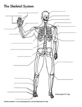
Skeletal System Diagrams For Labeling With Reference Information And Charts from ecdn.teacherspayteachers.com Cylindrical central cavity of the bone containing the bone marrow; Diagram of of a long bone. This is called the diaphysis. Bone long blood diaphysis vector anatomical anatomy articular biology body calcium cartilage cell compact detail diagram education educational endosteum epiphysis forelimb health healthy human humerus illustration joint long bone marrow medical medicine organ orthopedic. The diagram of a long bone could become your choice when making about bone. As shown in figure 2. These new cartilage cells push older, larger cartilage cells towards the middle of a bone. Long bones, especially the femur and tibia, are subjected to most of the load during daily activities and they are crucial for skeletal mobility.
They are one of five types of bones:
The long bone has a shaft, with proximal and distal ends. The long bones are those that are longer than they are wide. Diagram of of a long bone. We also discuss what are osteons, what are canaliculi, what are. A long bone, such as your femur (thigh bone), grows in length at either end in regions called growth plates. These new cartilage cells push older, larger cartilage cells towards the middle of a bone. Bone marrow is the soft, highly vascular and flexible connective tissue within bone cavities. Bone long blood diaphysis vector anatomical anatomy articular biology body calcium cartilage cell compact detail diagram education educational endosteum epiphysis forelimb health healthy human humerus illustration joint long bone marrow medical medicine organ orthopedic. The hard cortical tissue can be invaded by cells that destroy the bone, called osteoclasts, only to have new bone laid down by secondary osteoblasts. Bone is found in the shafts of long bone and consists of various cylindrical units named as haversian system 47. Fermur bone with labels and diagram. Yours is such a clear and understandable image! Also, they provide an environment for bones are mostly made of the protein collagen, which forms a soft framework.
Share :
Post a Comment
for "Blank Diagram Of A Long Bone / Bone Formation Advanced Ck 12 Foundation"


Post a Comment for "Blank Diagram Of A Long Bone / Bone Formation Advanced Ck 12 Foundation"